Segmental Anatomy: Sequential Segmental Analysis
Letter 1: atria (S, I or A)
The joint nomenclature statement (Jacobs 2007): heterotaxy, situs ambiguus and visceral heterotaxy all mean organs not clearly lateralised; by convention neither solitus nor inversus. The sequential school then sorts heterotaxy by the appendages, isomerism of the right or of the left atrial appendages; “atrial isomerism” does not exist, only the appendages are isomeric. Some morphologists, Van Praagh among them, do not accept isomerism of the appendages and write atrial situs ambiguus with the state of the spleen (Van Praagh 1984). Jacobs 2007 also lists X for unknown atrial situs.| Letter | Means, and how it shows | Source |
|---|---|---|
| Solitus: morphologically right atrium on the right; on subcostal, aorta left of the spine, IVC right | Jacobs 2007 | |
| Inversus: mirror image; aorta right, IVC left | Jacobs 2007 | |
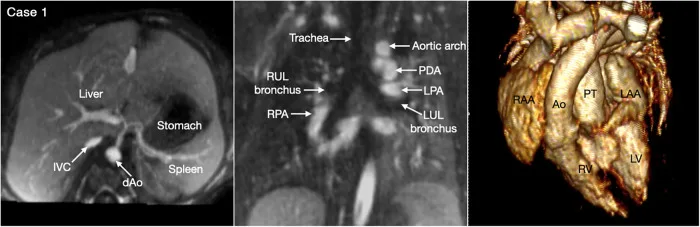
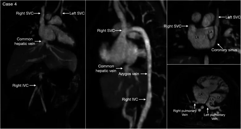
| Ambiguus (heterotaxy), in two forms.Right isomerism: both appendages right type; aorta and IVC almost always on one side, IVC slightly anterior.Left isomerism: both left type; IVC often interrupted, azygos or hemiazygos return: aorta midline, azygos posterolateral. | Jacobs 2007 |
How to tell
- The appendage names the atrium: right broad and blunt, pectinate muscles round the vestibule to the crux; left narrow, finger-like, pectinate muscles inside it only (Jacobs 2007; Anderson 2009). The IVC’s entry above the diaphragm is an extremely reliable marker of the right atrium only when the organs are lateralised; the abdominal vessel rule fails in heterotaxy (Jacobs 2007). Other atrial clues are not reliable: a left SVC can enter the left atrium, pulmonary veins can connect anomalously, an unroofed coronary sinus can make the IVC seem to enter the left atrium (Lai 3e Table 3.1). Appendages are hard to see in older children and adults: parasternal short axis, better on TOE (Bellsham-Revell 2019).
- Bronchi agree with the appendages more closely than the spleen does: right short and eparterial, left long and hyparterial (Jacobs 2007). On a chest radiograph, a ratio of less than 1 to 1.6 between the lengths of the two main bronchi is consistent with isomerism, more than 1 to 1.6 with lateralisation (Jacobs 2007, citing earlier work). The agreement with the appendages is close but not absolute.
- Not the veins or the spleen: describe them; they neither name the atria nor sort the heart disease (Jacobs 2007).
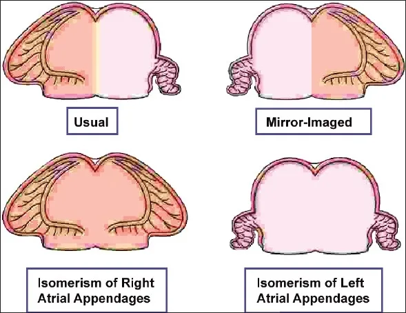
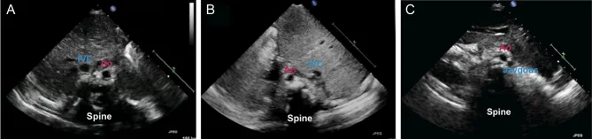
Right and left isomerism compared (Jacobs 2007)
| Feature | Isomeric right appendages | Isomeric left appendages |
|---|---|---|
| Lungs, bronchi | Often three lobes each, short bronchi | Often two lobes each, long bronchi |
| Spleen | Usually absent (asplenia) | Usually multiple (polysplenia) |
| Pulmonary veins | TAPVC always, even when all veins reach one atrium | Often to both atria |
| Coronary sinus | Always absent | Can be absent; an SVC draining to a coronary sinus is found only here |
| IVC | Not typically interrupted (asplenia syndrome) | Interruption with azygos or hemiazygos continuation is the commonest anomaly, not universal; in more than 80% (as cited in Yoo 2023) |
| Superior caval veins | Bilateral SVC frequent in both | |
| AV junction | Common junction in virtually all with asplenia syndrome; univentricular connection more frequent | Common junction in almost two-thirds with polysplenia syndrome |
| VA connection | Discordant or double outlet, often with severe pulmonary stenosis or atresia, more frequent | Concordant, with subaortic obstruction and coarctation, more frequent |
| Sinus node | Two sinoatrial nodes (Bellsham-Revell 2019) | No sinoatrial node; complete heart block can occur, ectopic atrial rhythm more usual (Bellsham-Revell 2019) |
| Gut | Malrotation frequent; some advise a barium study in all, above all with right isomerism | |
“Asplenia syndrome” and “polysplenia syndrome” are names for the usual pairing only: a right-isomerism patient may have a spleen, and spleen, lungs and appendages may disagree, so describe each. In some living patients the appendages cannot be settled at all; say so (Jacobs 2007).
Letter 2: ventricles (D, L or X)
| Letter | Means | Source |
|---|---|---|
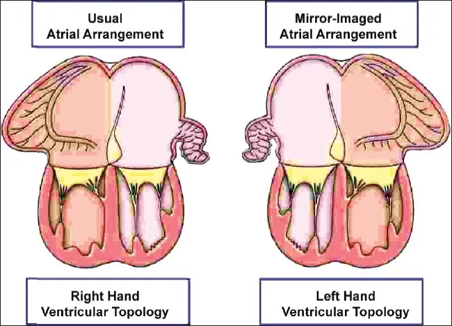
| D-loop (right-hand topology, normal): the right palm fits the septal surface of the morphologically right ventricle, thumb in the inlet, fingers in the outlet | IPCCC 2017; Anderson 2009 | |
| L-loop (left-hand topology): only the left hand fits | IPCCC 2017 | |
| Undetermined loop, allowed by many centres | Jacobs 2007 |
How to tell
- First name each ventricle by its own morphology (table below), never by which atrium or veins connect to it.
- Then the hand: it works wherever the ventricles lie; right-left position does not decide it (Lai 3e Ch 3 textbook). With the LV: a right-handed LV means an L-loop, a left-handed LV a D-loop. The loop cannot be decided in an anatomically single RV with no recognisable LV or septum (Lai 3e Fig 3.10). On 3D imaging of a distorted heart, turn the ventricles to the “ventricular apical view”: septum vertical, apex toward you, arteries up (Yoo 2023).
- Diagnose the loop itself, not from the AV connections; it sets the coronary pattern and conduction system (Van Praagh 1984). Van Praagh 1984: the loop cannot be inferred from the AV alignments with consistent accuracy. Lai 3e: an L-loop is associated with AV block, an Ebstein-like left-sided tricuspid valve and a hypoplastic left-sided RV sinus.
| Ventricle | Its own marker | Source |
|---|---|---|
| Right ventricle | Coarse apex; tricuspid cords on the septum; moderator band; more apical AV valve Sources differ on the single best sign: apical trabeculations (Anderson 2009), the smooth superior septal surface of the LV (Lai 3e), apical offset of the right-sided AV valve (Bellsham-Revell 2019; reverse offset suggests discordant AV connections). Hypertrophy or double inlet can coarsen LV trabeculations (Lai 3e). When one ventricle is hypoplastic, position helps on imaging: a small LV almost always lies inferiorly by the crux, an incomplete RV almost always anterior and superior (Yoo 2023). | Anderson 2009; Bellsham-Revell 2019 |
| Left ventricle | Fine apex; smooth superior septal surface; papillary muscles on the free wall only | Lai 3e Ch 3 textbook |
| Indeterminate | Very coarse apex with large muscle bundles; no second ventricle | Anderson 2009 |
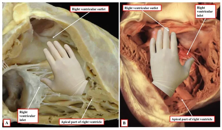
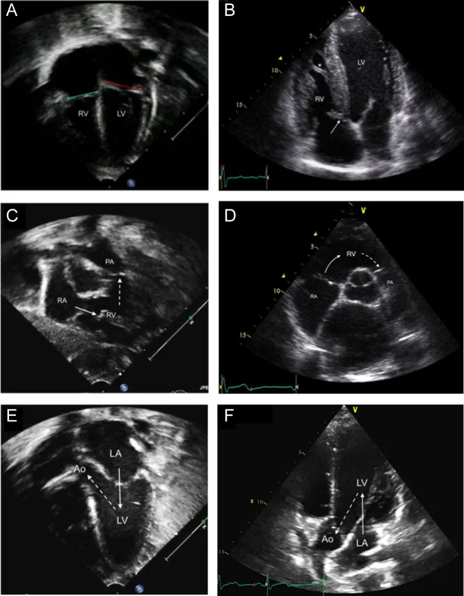
Letter 3: great arteries (S, I, D, L or A)
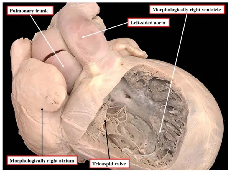
The five letters and their meanings as defined in the joint nomenclature statement (Jacobs 2007): S normally related, I inverted (mirror-image) normally related, D D-transposed or D-malposed, aorta right of the pulmonary trunk, L L-transposed or L-malposed, aorta left, A aorta directly anterior. Crucean 2022 lists S, I, D and L, with D and L the two options for an abnormal location of the aortic root. “L-TGA always means” the aortic valve left of the pulmonary valve (Van Praagh 1984). A: IPCCC 2017 “A-malposed aorta”, aortic root directly anterior (02.06.03), and Lai 3e Fig 3.14. IPCCC codes: posterior and rightward 02.06.00 (synonym “normally related great arteries”), posterior and leftward 02.06.06 (“mirror imaged normally related”).| Letter | Aorta relative to the pulmonary trunk | Source |
|---|---|---|
| Normally related: aortic valve posterior and to the right of the pulmonary valve | Jacobs 2007; IPCCC 2017 | |
| Inverted normal (mirror image): posterior and to the left | Jacobs 2007; IPCCC 2017 | |
| Abnormally related (transposed or malposed), aorta to the right | Jacobs 2007 | |
| Abnormally related (transposed or malposed), aorta to the left | Jacobs 2007; Van Praagh 1984 | |
| Abnormally related, aorta directly anterior | Jacobs 2007; IPCCC 2017 |
Anterior
← Patient’s rightPatient’s left →
in front, to the right
directly in front
in front, to the left
beside, to the right
Pulmonary
valve
valve
beside, to the left
behind, to the right: normally related
behind, to the left: mirror-image normal
Posterior
The rest of the analysis
Lai 3e lists 10 steps: situs, position, segment by segment, atria, loop, AV, VA, infundibulum, semilunar relations, associated anomalies; Yoo 2023 lists 5. The three letters are steps 4, 5 and 9. The rules: name each segment by its own morphology; state situs and connections separately, never inferring one from the other (Lai 3e Fig 3.2); keep morphology, connections and relations apart (Anderson 2009).Situs: bronchi, liver and spleen; position and apex
Abdominal organs, bronchi and atria each on its own: about 20% of abnormal situs disagrees across the three (as cited in Yoo 2023).
| Right: 3 lobes, short eparterial bronchus. Left: 2 lobes, long hyparterial bronchus | Lai 3e Ch 3 textbook; Jacobs 2007 | |
| Right: 2 lobes, hyparterial. Left: 3 lobes, eparterial | Lai 3e Ch 3 textbook | |
| Both lungs 3 lobes, both bronchi short and eparterial; often with right isomerism | Jacobs 2007; Lai 3e Ch 3 textbook | |
| Both lungs 2 lobes, both bronchi long and hyparterial; often with left isomerism | Jacobs 2007; Lai 3e Ch 3 textbook |
| Usual arrangement | Lai 3e Ch 3 textbook | |
| Mirror image | Lai 3e Ch 3 textbook | |
| Seen in both isomerisms | Bellsham-Revell 2019; Lai 3e Ch 3 textbook |
| Usual arrangement | Lai 3e Ch 3 textbook | |
| Mirror image; also among the splenic anomalies of heterotaxy | Lai 3e Ch 3 textbook | |
| Right isomerism usually has this | Jacobs 2007 | |
| Left isomerism usually has this | Jacobs 2007 |
| Heart mostly in the left chest | IPCCC 2017 | |
| Midline | IPCCC 2017 | |
| Mostly in the right chest; a position, not a diagnosis Dextrocardia usually means a right-sided ventricular mass, dextroversion an apex pointing right; a right-chest heart with a leftward apex is “dextrocardia with levoversion”. Avoid compressed terms such as pivotal or mixed dextrocardia; position and apex matter for an extracardiac Fontan route (Jacobs 2007). Primary dextrocardia comes with a structural heart defect, apex usually to the right. Secondary: pushed (left tension pneumothorax, left congenital lobar emphysema, left diaphragmatic hernia) or pulled (right lung hypoplasia or agenesis); the apex may point left or forward (Lai 3e). A right-sided heart may also be pulled right, as in Scimitar syndrome (Bellsham-Revell 2019). | IPCCC 2017 |
| Judged apart from the position | Jacobs 2007 | |
| Jacobs 2007 | ||
| Jacobs 2007 |
Veins
| SVC and IVC to the right atrium, four pulmonary veins to the left atrium | Bellsham-Revell 2019 | |
| Frequent in both isomerisms | Jacobs 2007 | |
| Among heterotaxy, only with left isomerism | Jacobs 2007 | |
| Azygos or hemiazygos continuation; the commonest anomaly in left isomerism | Jacobs 2007 | |
| Always present with right isomerism | Jacobs 2007 |
Atrioventricular connection
| RA to RV, LA to LV | Anderson 2009 | |
| RA to LV, LA to RV | Anderson 2009 | |
| Isomeric appendages, two ventricles; state the topology With isomeric appendages and two atria joined to two ventricles, half the heart is joined concordantly and half discordantly, whatever the topology. The European code called this “ambiguous”; “mixed” is the better word and is now in the IPCCC (Jacobs 2007). | Anderson 2009; Jacobs 2007 | |
| Both atria to one ventricle | Anderson 2009 | |
| Right-sided atrium not joined to a ventricle Atresia is usually an absent connection (fibro-fatty groove between atrium and ventricle); an imperforate valve is atresia with a potential connection (Anderson 2009). Van Praagh calls the first simply tricuspid or mitral atresia and the second an imperforate valve, and finds “with absent AV connection” redundant (Van Praagh 1984). | Anderson 2009 | |
| Left-sided atrium not joined to a ventricle | Anderson 2009 |
| Anderson 2009 | ||
| Anderson 2009 | ||
| Anderson 2009 |
| Anderson 2009 | ||
| Anderson 2009 | ||
| Anderson 2009 | ||
| Cords on both sides of the septum | Anderson 2009 | |
| Annulus over the septum | Anderson 2009 |
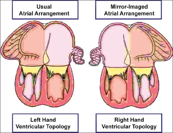
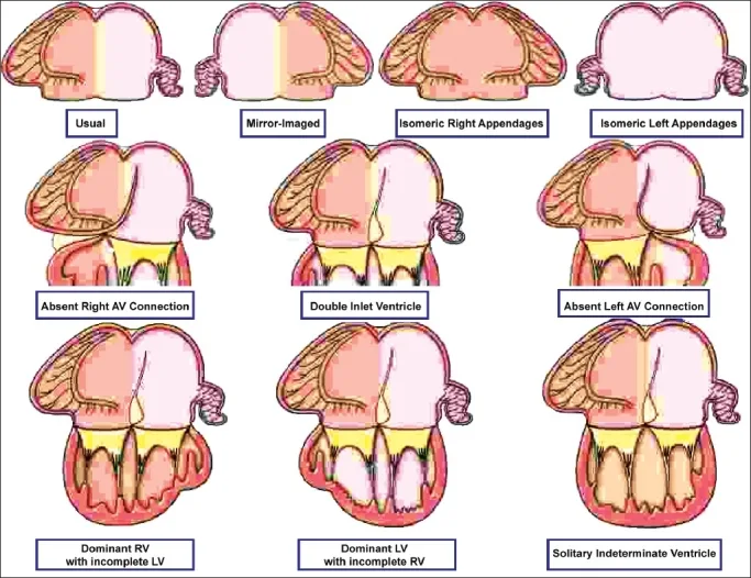
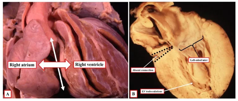
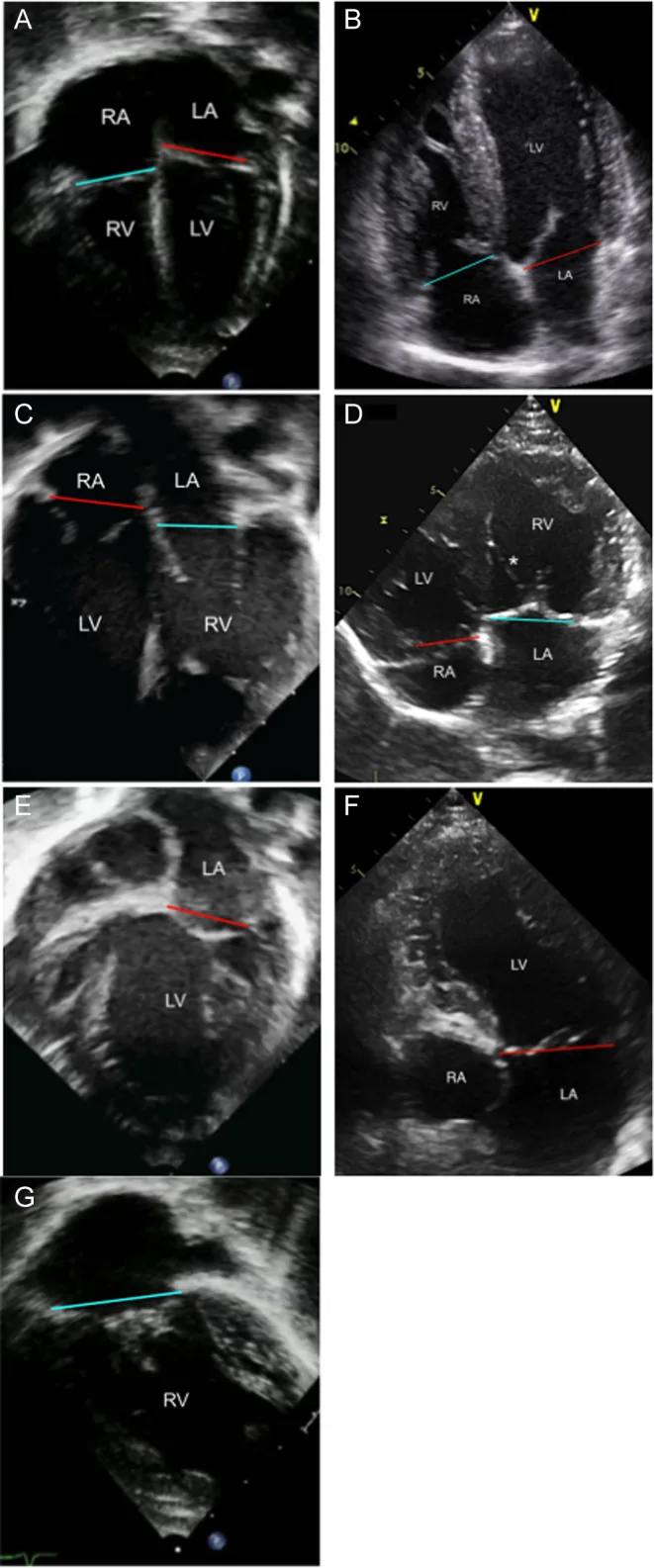
Ventriculo-arterial connection
| Aorta from the LV, pulmonary trunk from the RV | Anderson 2009 | |
| Aorta from the RV, pulmonary trunk from the LV | Anderson 2009 | |
| Both arteries from the RV | Anderson 2009 | |
| Both arteries from the LV | Anderson 2009 | |
| Single outlet with one valve and one trunk Single outlet: common trunk; solitary trunk (no pulmonary trunk remnant found); pulmonary trunk with aortic atresia, or aortic trunk with pulmonary atresia, when the atretic vessel’s ventricle cannot be shown (Anderson 2009). An overriding arterial valve is given to the ventricle supporting most of its circumference (Anderson 2009); Lai 3e finds the “50% rule” not practicable in vivo and describes the anatomy instead (e.g. RV origin of the aorta, biventricular origin of the pulmonary artery). | Anderson 2009 | |
| Single outlet, no pulmonary trunk remnant | Anderson 2009 | |
| Single outlet, only when the atretic aorta’s ventricle cannot be shown; otherwise pick the connection and an imperforate valve | Anderson 2009 | |
| Single outlet, only when the atretic pulmonary trunk’s ventricle cannot be shown; otherwise pick the connection and an imperforate valve | Anderson 2009 |
| One or both may override | Anderson 2009 | |
| The connection is still stated, with the imperforate valve | Anderson 2009 |
| Normal | Lai 3e Ch 3 textbook | |
| Often transposition | Lai 3e Ch 3 textbook | |
| Often DORV | Lai 3e Ch 3 textbook | |
| Rare; most often DOLV | Lai 3e Ch 3 textbook; Anderson 2009 |
On echo normal arteries cross; in transposition they usually run parallel (Bellsham-Revell 2019).
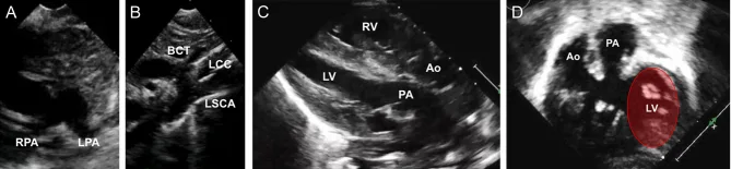
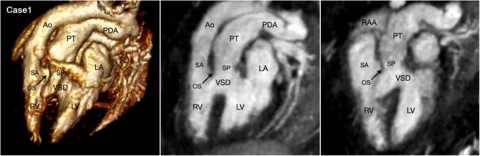
All the variables at a glance
The variables at a glance (Anderson 2009 flow diagram)
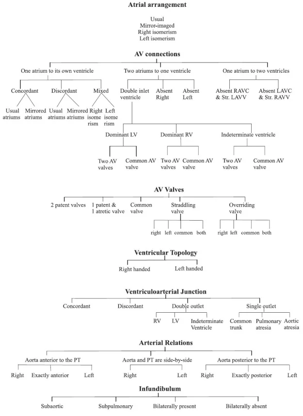
How to read and write the three letters of a segmental set, then the rest of the analysis, in Van Praagh, sequential and IPCCC terms. Terms and method only; no values.
Sources and how this page was checked
Full text read
- Jacobs JP, Anderson RH, Weinberg PM, et al. The nomenclature, definition and classification of cardiac structures in the setting of heterotaxy. Cardiol Young 2007;17(Suppl 2):1-28. doi:10.1017/S1047951107001138. Full text (publisher PDF, 28 pp). The joint statement of the Nomenclature Working Group, Boston and European authors together: the notation letters including X, the definitions of heterotaxy, isomerism and situs ambiguus, the atrial, venous and junctional features of the two isomerisms, and the position terms. No figure reproduced.
- Van Praagh R. The segmental approach clarified [letter]. Cardiovasc Intervent Radiol 1984;7(6):320-325. doi:10.1007/BF02625121. Publisher scan. Alignments and connections, the Kirklin and London modifications, D- and L-loops, isomerism, the loop rule, braces and sets.
- Anderson RH, Shirali G. Sequential segmental analysis. Ann Pediatr Cardiol 2009;2(1):24-35. doi:10.4103/0974-2069.52803. Open access, PMC2840778; figures under CC BY 2.0. review
- Crucean AC, Spicer DE, Anderson RH. The significance of ventricular topology in the analysis of congenitally malformed hearts. J Cardiovasc Dev Dis 2022;9(5):155. doi:10.3390/jcdd9050155. Open access, PMC9144843; CC BY 4.0. A sequential-school account of the notation; source of the TGA {S,D,L}, DORV {S,D,L} and DILV {S,D,D} examples. review
- Franklin RCG, Béland MJ, Colan SD, et al. Nomenclature for congenital and paediatric cardiac disease: the International Paediatric and Congenital Cardiac Code (IPCCC) and the Eleventh Iteration of the International Classification of Diseases (ICD-11). Cardiol Young 2017;27(10):1872-1938. doi:10.1017/S1047951117002244. Table 2, pp 1900-1903, read as page images.
- Jacobs JP, Franklin RCG, Béland MJ, et al. Nomenclature for pediatric and congenital cardiac care: unification of clinical and administrative nomenclature. Cardiol Young 2021;31(7):1057-1188. doi:10.1017/S104795112100281X. Checked that it repeats the chirality and heterotaxy entries.
- Bellsham-Revell H, Masani N. Educational series in congenital heart disease: the sequential segmental approach to assessment. Echo Res Pract 2019;6(1):R1-R8. doi:10.1530/ERP-17-0039. Open access, PMC6301191; figures shown whole and unchanged under CC BY-NC-ND 4.0. review
- Yoo SJ, Perens G, Nguyen KL, et al. Contemporary sequential segmental approach to congenital heart disease using four-dimensional MR angiography. Front Cardiovasc Med 2023;10:1107399. doi:10.3389/fcvm.2023.1107399. Open access, PMC10352920; CC BY. Its percentages are cited from earlier work, not its own series. Its Figure 4 was published before and is not used. review
- Geva T. Segmental approach to congenital heart disease. Ch 3 in: Lai WW, Mertens LL, Cohen MS, Geva T, eds. Echocardiography in Pediatric and Congenital Heart Disease, 3rd ed. Wiley; 2021. doi:10.1002/9781119612858. Chapter text, Table 3.1 and Figures 3.2, 3.3, 3.4, 3.10, 3.12, 3.13 and 3.14 read; no textbook figure is reproduced. textbook
Not read for this page
- Van Praagh’s original papers: Am J Cardiol 1964;13:510-531 (cited in Van Praagh 1984), Birth Defects 1972;8:4-23, and Cardiovasc Intervent Radiol 1984;7:115-120 (the paper the 1984 letter answers). The letters rest on Jacobs 2007 and Crucean 2022 and the examples in Van Praagh 1984; they agree.
- Shinebourne EA, et al. Br Heart J 1976 and Tynan MJ, et al. Br Heart J 1979 (the London papers), described only as Van Praagh 1984 and Anderson 2009 describe them.
How it was checked
- Van Praagh’s segmental approach and Anderson’s sequential segmental analysis are, in Van Praagh’s words, the same system “expressed in different words”. He calls what opens into what an alignment and keeps connection for the junctional segments, and holds that concordant and discordant do not apply to tricuspid atresia, double inlet, straddling valves or situs ambiguus; Crucean 2022 reads the original sets as implying AV concordance for {S,D,*} or {I,L,*} whatever the connection. The loop rule has many exceptions (Van Praagh 1984).
- Terms are the sources’ own, condensed (November 2026); no number on this page drives a decision. Where sources disagree (the best single sign of a ventricle; isomerism or situs ambiguus; the 50% rule; whether concordance applies to double inlet), each view is given with its source.
- The letters and their meanings follow Jacobs 2007, which also allows X for unknown atrial situs and, in many centres, an undetermined loop. Crucean 2022 lists S, I, D and L for the arterial segment; A for a directly anterior aorta is in Jacobs 2007, IPCCC 2017 and Lai 3e.
- Figures are reproduced whole from open-licence papers, each credited under it; no figure was redrawn or changed.
- As printed: IPCCC 2017 lists “Aortic root posterior and rightward to pulmonary root (02.06.06)” among its normal terms (p 1881), but Table 2 gives 02.06.06 to posterior and leftward and 02.06.00 to posterior and rightward; this page uses the Table 2 codes. Bellsham-Revell 2019 Table 1 prints “midline liver” for both isomerisms. Its TAPVC “by definition” in right isomerism agrees with Jacobs 2007: with no morphologically left atrium, pulmonary venous connection is anomalous even when all veins reach one atrium.
- Source extract:
verification/sources/segmental-anatomy.md. Independent review in three rounds before release.